
Fig. 4
- ID
- ZDB-FIG-140107-57
- Publication
- Borovina et al., 2013 - IFT88 Plays a Cilia- and PCP-Independent Role in Controlling Oriented Cell Divisions during Vertebrate Embryonic Development
- Other Figures
- All Figure Page
- Back to All Figure Page
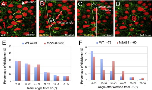
IFT88 Is Required for PCP-Independent OCD at Neurulation (A?D) Confocal images of neural progenitor cell division in a six-somite-stage (12 hpf) WT embryo expressing Arl13b-GFP (green; cell membranes) and histone 2B-Cherry (red; histones). In these cells, the initial orientation of the metaphase plate is realigned to be parallel to the midline so that the subsequent division plane occurs across the midline. Images depict a cell that is about to undergo cell division (A), the initial orientation of the metaphase plate (B), metaphase plate orientation following spindle rotation (C), and the cell in anaphase (D). Elapsed time (t) is indicated in minutes. Spindle orientation was measured as the angle of the metaphase plate relative to the neural midline. (E) Graph of spindle orientations at the onset of metaphase, demonstrating no significant difference (p = 0.830) between WT (blue; n = 73) and MZift88 (red; n = 60) cells. (F) Graph of metaphase plate orientations following spindle rotation. The metaphase plate in WT cells (blue; n = 73) is strongly aligned with the midline, whereas MZift88 cells (red; n = 60) exhibit a statistically significant shift (p = 0.010) to a more randomized orientation. Statistical significance was determined using a Watson-Williams two-sample test and statistical significance was assigned when p < 0.05. |
| Fish: | |
|---|---|
| Observed In: | |
| Stage: | 5-9 somites |