
Fig. 2
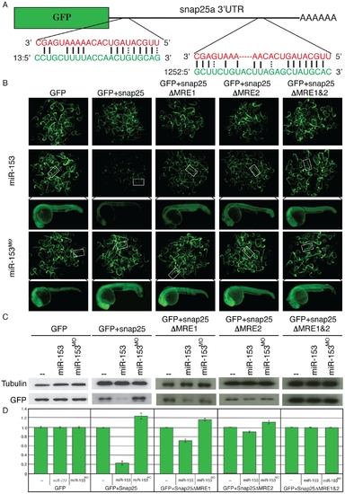
miR-153 targets snap-25a. (A) GFP reporter constructs were created by fusing the reading frame of GFP to the snap-25a 32UTR. Two predicted miRNA recognition elements (MREs) were identified in the snap-25a 32 UTR. The miR-153 sequence is indicated in red and the corresponding snap-25a UTR sequence is shown in green. (B) Single cell zebrafish embryos were injected with mRNAs derived from GFP reporters lacking a UTR (GFP), fused to the full length snap-25a UTR (+snap-25), or mutant versions of the snap-25a UTR lacking individual MREs (snap-25aΔMRE1 and snap-25a?MRE2) or both MREs (snap-25aΔMRE1&2). Embryos were injected in the presence or absence of exogenous miR-153 or morpholinos against miR-153 (miR-153MO). Fluorescence levels were examined at 1 dpf. Clusters of embryos (~60) are shown as well as a high magnification image of a single representative embryo. (C) Lysates from ~100 embryos were prepared from embryos treated as in B and GFP protein levels were determined by western blotting using antibodies against GFP or control antibodies against α-tubulin. (D) Quantitation of westerns was performed with a paired Student?s t-test (n = 5). |