
FIGURE
Fig. 3
- ID
- ZDB-FIG-130910-22
- Publication
- Kim et al., 2013 - Multi-organ Abnormalities and mTORC1 Activation in Zebrafish Model of Multiple Acyl-CoA Dehydrogenase Deficiency
- Other Figures
- All Figure Page
- Back to All Figure Page
Fig. 3
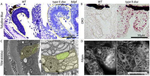
Kidney defects in 8 dpf dxavu463 mutant zebrafish. (A) Toluidine blue staining in pre-TEM sections at 8 dpf. Various sized vacuoles were observed in the dxa mutants. (B) ORO staining showing marked increase in lipids in dxa mutants. (C) TEM of kidney epithelium in wild type (left) and dxa mutants (right). Green colored regions indicate rod-shaped mitochondria in the kidney. (D) Filipin staining again shows free cholesterol accumulation in the cytosol of kidney cells. N, nuclei. Scale bar = 50 μm (A, B, D) and 2 μm (C). |
Expression Data
Expression Detail
Antibody Labeling
Phenotype Data
| Fish: | |
|---|---|
| Observed In: | |
| Stage: | Days 7-13 |
Phenotype Detail
Acknowledgments
This image is the copyrighted work of the attributed author or publisher, and
ZFIN has permission only to display this image to its users.
Additional permissions should be obtained from the applicable author or publisher of the image.
Full text @ PLoS Genet.