Fig. 6
- ID
- ZDB-FIG-130905-17
- Publication
- Filipek-Górniok et al., 2013 - Expression of chondroitin/dermatan sulfate glycosyltransferases during early zebrafish development
- Other Figures
- All Figure Page
- Back to All Figure Page
|
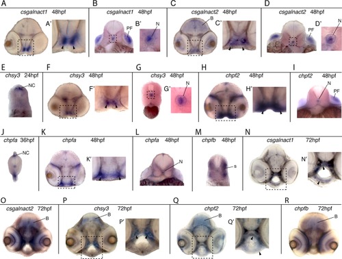
Sections of embryos subjected to in situ hybridization of CS/DS glycosyltransferases. Transversal sections showing the expression patterns of csgalnact1 at 48 hpf (A,B) and 72 hpf (N,O), csgalnact2 at 48 hpf (C,D) and 72 hpf (P,Q), chsy3 at 24 hpf (E), 48 hpf (F,G) and 72 hpf (R,S), chpf2 at 48 hpf (H,I) and 72 hpf (T), chpfa at 36 hpf (J) and 48 hpf (K,L), and chpfb at 48 hpf (M) and 72 hpf (U). The position of each section is indicated with an arrow marked in Figure 4 and figure 5. Magnifications of boxed areas are shown to the right (A–D, F–H, K, N, P, Q). Arrowheads mark pharyngeal cartilage. B, brain; N, notochord; NC, neural crest; PF, pectoral fin. |
| Genes: | |
|---|---|
| Fish: | |
| Anatomical Terms: | |
| Stage Range: | Prim-5 to Protruding-mouth |

