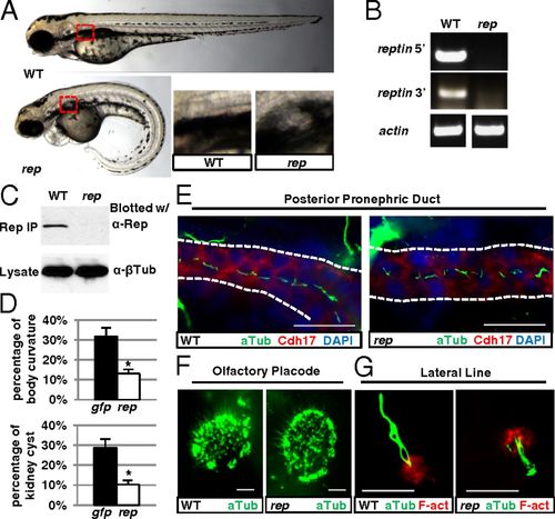
Phenotypes of reptinhi2394 mutants. (A) Reptinhi2394 mutant (rep) at 3 d postfertilization (dpf) showing kidney cyst (red box, magnified in Lower Right) and body curvature, compared with wild-type (WT) fish. (B) RT-PCR using lysates of 2-dpf reptinhi2394 (rep) and WT siblings. Two pairs of primers, one to the 52 side of the proviral insertion and one to the 32 side, were used. (C) The greatly reduced Reptin protein in 4-dpf reptinhi2394 mutant (rep). (Upper) Samples precipitated and blotted with anti-Reptin. (Lower) Lysates blotted with anti?β-tubulin as a loading control. (D) Microinjection of reptin-eGFP mRNA (rep) reduced the percentages of body curvature (Upper) and kidney cysts (Lower) in embryos from reptinhi2394+/ crosses, with eGFP mRNA (gfp) microinjection as a negative control. Data are represented as mean + SD, from three replicates. *P < 0.05. (E?G) Immunostaining showing cilia (in green) in reptinhi2394 mutants (rep) in the posterior pronephric duct (E) (stained with anti-Cdh17, a marker for kidney epithelial cells, in red and lined with dotted lines), the olfactory placode (F), and the lateral line organ (G) [F-actin (F-act) stained with rhodamine phalloidin in red]. (Scale bar, 20 μm in E and F and 10 μm in G.)
|
