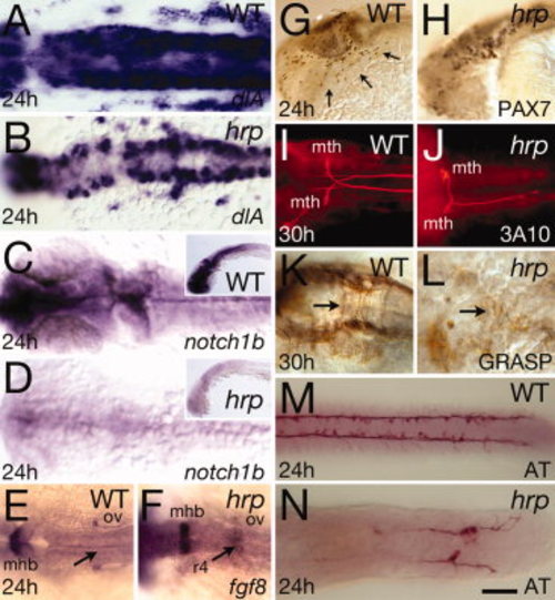
Neural patterning defects in harpy mutants. Panels show wild-type control embryos and harpy mutants. A,B: Expression of deltaA (dlA) at 24 hr. Dorsal view of hindbrain. Note the normal intensity of expression in mutant cells. C,D: Expression of notch1b at 24 hr. Dorsal view of head; insets show lateral view. Note expression in mutants is almost absent. E,F: Expression of fgf8 at 24 hr. Dorsal view of hindbrain. Note ectopic staining in the fourth rhombomere of the mutant. G,H: Anti-Pax7 staining at 24 hr. Arrows indicate neural crest cells migrating over the eye and pharyngeal arches; while these cells are present in the mutant, they are not seen on the migration pathway. I,J: Dorsal view of 3A10-stained Mauthner neurons at 30 hr, displaying abnormal pathfinding and absence of axons in the mutants. K,L: Anti-Grasp staining at 30 hr; dorsal view of the hindbrain. Arrows indicate commissural neurons that in the mutant are chaotic. M,N: Anti-acetylated tubulin (AT) staining at 24 hr; dorsal view of the trunk. Although axons are present in mutants, they do not express acetylated tubulin except in the tail region. mhb, midbrain?hindbrain border; mth, Mauthner neurons; ov, otic vesicle; r4, fourth rhombomere. Scale bar = 50 μm in A?D, 20 μm in F,G, 100 μm in E?N.
|
