
Fig. 2
- ID
- ZDB-FIG-130426-21
- Publication
- O'Reilly-Pol et al., 2013 - Kit signaling is involved in melanocyte stem cell fate decisions in zebrafish embryos
- Other Figures
- All Figure Page
- Back to All Figure Page
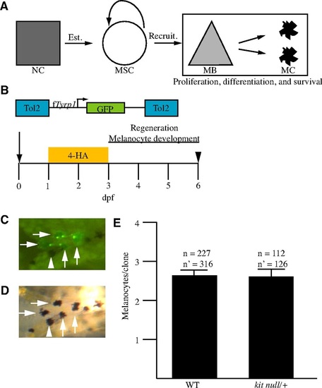
MSC daughter cell proliferation, differentiation and survival are unaffected in kitanull/+ embryos. (A) A general model of melanocyte regeneration. A neural crest progenitor (square, NC) establishes a melanocyte stem cell (circle, MSC), which can be recruited to produce melanoblasts (triangle, MB), which will proliferate and differentiate into melanocytes (MC). Box indicates process tested. (B) Schema of the experiment. Zebrafish embryos were injected at the one- to two-cell stage (arrow) with transposase and a transposon containing fTyrp1>GFP, treated with 4-HA from 1-3 dpf to ablate ontogenetic melanocytes, and scored for regeneration melanocytes at day 6 (arrowhead). (C,D) Example of a clone in WT. (C) An epifluorescent image of an embryo containing a clone. (D) A brightfield image of the embryo shown in C. Melanocytes containing GFP are marked with an arrow. Some melanocytes lack GFP, and one example is marked with an arrowhead. (E) The mean number of melanocytes per clone is the same for both genotypes in regeneration. n, the number of embryos containing clones; n′ the number of clones after adjustment for polyclonal events. Error bars are s.e.m. |