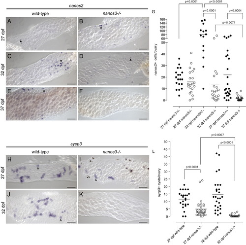
nanos2+ and sycp3+ cells are not maintained in nanos3 mutant juvenile ovaries. (A?G) Analysis of nanos2-expressing cells by in situ hybridization in wild-type and nanos3 mutant 27?37 dpf ovaries. nanos2+ cells are easily detected in wild-type and mutant ovaries at 27 dpf. (A and B). At 32 dpf, nanos2+ cells are easily detectable in both wild-type and mutant ovaries, but are more abundant in wild-type (C-D). However, by 37 dpf nanos2+ cells are not detected in mutants, but are present in wild-type ovaries (E and F) Arrowheads identify representative nanos2-expressing cells in A?E. (G) Quantification of nanos2+ cells per ovary in wild-type and mutant ovaries. (H?L) Analysis of sycp3 expressing germ cell cysts by in situ hybridization in wild-type and mutant ovaries from 27?37 dpf. Actively differentiating sycp3 expressing germ cell cysts are present in both wild-type and mutant tissue at 27 dpf (H and I). By 32 dpf sycp3+ cysts cannot be detected in mutant ovaries (J and K). Arrowheads identify representative sycp3 expressing germ cell cysts (blue) in H?J, while an asterisk in I identifies a representative melanocyte pigment cell (black). (L) Quantification of sycp3+ cysts per ovary in wild-type and mutant ovaries. All images are from whole mount preparations. Scale bars: 50 μm. p-values indicate statistically significant differences.
|
