
FIGURE
Fig. 6
- ID
- ZDB-FIG-130208-22
- Publication
- Monk et al., 2013 - Mutation of sec63 in zebrafish causes defects in myelinated axons and liver pathology
- Other Figures
- All Figure Page
- Back to All Figure Page
Fig. 6
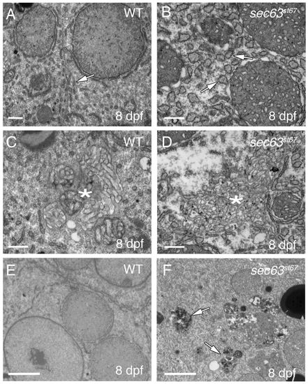
sec63st67 mutants develop numerous liver pathologies. (A–F) TEM images showing liver ultrastructure at 8 dpf in wild-type and sec63st67 mutant zebrafish. (A) Wild-type liver; arrow points to ER with normal morphology. (B) sec63st67 mutant liver; ER is swollen (arrows) and cytoplasm is disrupted. (C,D) Ultrastructure of bile canaliculi (*) in wild-type (C) and sec63st67 mutant (D) livers. sec63st67 bile canaliculi appear disorganized compared with the wild type. Lysosomes are filled with debris in sec63st67 mutants (F; arrows) but not in the wild type (E). Scale bars: 500 nm (A–D); 2 μm (E,F). |
Expression Data
Expression Detail
Antibody Labeling
Phenotype Data
| Fish: | |
|---|---|
| Observed In: | |
| Stage: | Days 7-13 |
Phenotype Detail
Acknowledgments
This image is the copyrighted work of the attributed author or publisher, and
ZFIN has permission only to display this image to its users.
Additional permissions should be obtained from the applicable author or publisher of the image.
Full text @ Dis. Model. Mech.