Fig. 5
- ID
- ZDB-FIG-130129-14
- Publication
- Folgueira et al., 2012 - Morphogenesis underlying the development of the everted teleost telencephalon
- Other Figures
- All Figure Page
- Back to All Figure Page
|
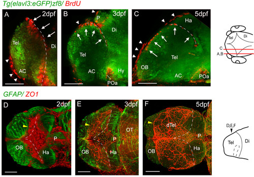
Proliferative cells and tela choroidea expand over the upper telencephalic surface between 2 dpf and 5 dpf. A, B and C. Parasagittal sections of BrdU-labelled (red) telencephali at 2, 3, and 5 days. At 2 days, the BrdU nuclei in the telencephalon (arrows) are seen lining the posterior telencephalic wall. By 3 days, a few nuclei are found on the upper surface of the telencephalon, and by 5 days, a more extensive area of the upper surface is lined with BrdU-positive nuclei (arrows). The more flattened BrdU-positive nuclei (arrowheads) are present in the overlying skin. D, E and F. ZO1 staining (red) reveals that the tela choroidea spreads over the dorsal surface of telencephalon over the same period as the proliferative cells. The diamond-shaped roof of the TDR at 2 dpf expands (yellow arrowhead) to cover the upper telencephalic surface (except for the OBs) by 5 dpf. Each image is a dorsal view created by projecting a z-stack of horizontal confocal sections. Neuroepithelium is counterstained with an anti-GFAP antibody (green). Dotted lines mark the position of the posterior telencephalic wall. Schematics on the right show levels of sections and planes of view. Scale bars 50 μm. AC: anterior commissure; AIS: anterior intraencephalic sulcus; Di: diencephalon; dTel: dorsal telencephalon; Hab: habenula; OB: olfactory bulb; P: pineal; POa: preoptic area; Tel: telencephalon. Dashed lines indicate medial and posterior walls of the telencephalon. |

