Fig. 7
- ID
- ZDB-FIG-121119-8
- Publication
- Rodriguez-Mari et al., 2011 - Roles of brca2 (fancd1) in Oocyte Nuclear Architecture, Gametogenesis, Gonad Tumors, and Genome Stability in Zebrafish
- Other Figures
- All Figure Page
- Back to All Figure Page
|
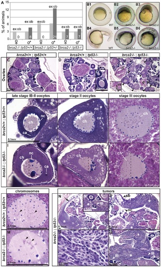
Mutation of tp53 rescued the brca2 sex reversal phenotype, yielding infertile double-mutant females that produced ovaries containing oocytes with altered nuclear architecture that developed into defective embryos and that produced invasive ovarian tumors. (A) Expected (ex) and observed (ob) frequencies of various tp53 genotypes among the homozygous brca2 mutant offspring of parents doubly heterozygous for tp53M214K and brca2 ZM_00075660 alleles. Females developed only in the absence of a wild-type tp53 allele, showing that the tp53 mutation rescued the sex reversal phenotype. (B) Doubly heterozygous embryos from the mating of homozygous tp53 females to males heterozygous for the brca2 mutation develop like normal embryos (B1 and B4 at 6 and 19 hpf, respectively) but the same genotype developing from the mating of rescued brca2;tp53 double mutant females and homozygous wild-type males stopped developing shortly after gastrulation (B2, B3, B5, B6). (C-E) Sections of ovaries from 6mpf wild-type, single mutant tp53, and double mutant brca2;tp53 adults, respectively, showed that oocytes in tp53 mutants developed normally (D′) but that oocytes degenerated and the vitelline envelope was abnormally separated from the granulosa cells in double mutants (E′). Strikingly, oocytes in double mutants showed nuclear anomalies (arrowheads in E). (F-H) Sections of wild-type late stage IB (transitioning to stage II), stage II, and stage III oocytes, respectively, contained radially symmetrical nuclei with small round nucleoli located mostly near the nuclear envelope and thin chromosomes separated from each other in the middle of the nucleus. (I-K) Sections of late stage IB, stage II, and stage III oocytes of brca2;tp53 double mutants showed abnormal nuclear asymmetry. Oocytes contained nuclei with large, variably-shaped nucleoli clustered at one side of the periphery of the nucleus instead of the normal uniform distribution around the periphery of the nucleus and chromosomes clustered at the other side of the nucleus opposite to the nucleoli. (L) Chromosomes in wild types were thin and separated (arrowheads). (M) Chromosomes in oocytes of double mutants were thicker and clumped and formed abnormal crosses and loops (arrowheads). (N,P) Invasive ovarian tumors (asterisks) were detected in sections of two of four brca2;tp53 double mutants by 6mpf but were not present in other genotypes (wild-types and single tp53 mutants). (N) One tumor appeared to originate at the ovarian cavity membrane (arrow) and the other tumor (P) was metastatic and contacted the ovarian cavity membrane and the swim bladder. (N′,Q) Both tumors were formed by spindle-shaped cells (O) that invaded the ovary and surrounded the oocytes. Abbreviations: IB,II,III: oocyte stages; ca, cortical alveoli; ch, chromosomes; d, degenerating oocyte; gc, granulosa cell; nc, nucleoli; oc, ovarian cavity; sb, swim bladder; ve, vitelline envelope. |
| Fish: | |
|---|---|
| Observed In: | |
| Stage: | Adult |

