
Fig. S2
- ID
- ZDB-FIG-121101-29
- Publication
- Cox et al., 2012 - An essential role of variant histone h3.3 for ectomesenchyme potential of the cranial neural crest
- Other Figures
- All Figure Page
- Back to All Figure Page
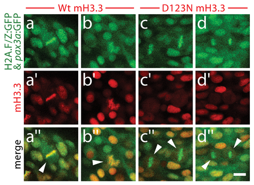
D123N H3.3 fails to localize to condensed chromosomes within NPB cells. Confocal images of 13 hpf embryos harboring the H2A.F/Z:GFP and NPB-specific pax3a:GFP transgenes and injected with wild-type or D123N versions of mCherry(m)H3.3 fusion proteins. a?d, GFP fluorescence of cells within the pax3a:GFP-labeled NPB domain. Whereas most cells are in interphase, the few cells in metaphase/anaphase (arrowheads in merged images: a′′?d′′) exhibit both GFP-labeled condensed chromosomes (H2A.F/Z:GFP) and more diffuse lower-level cytoplasmic GFP (pax3a:GFP). a2?b2, Wild-type mCherry-H3.3 localizes within the condensed chromosomes of 14/14 metaphase/anaphase cells. c′?d′, D123N mCherry-H3.3 fails to localize within condensed chromosomes and instead appears diffuse throughout pax3a:GFP-positive NPB cells after nuclear envelope breakdown. (0/14 cells exhibit chromosomal localization during metaphase/anaphase). Scale bar = 10 μm. |