
Fig. 5
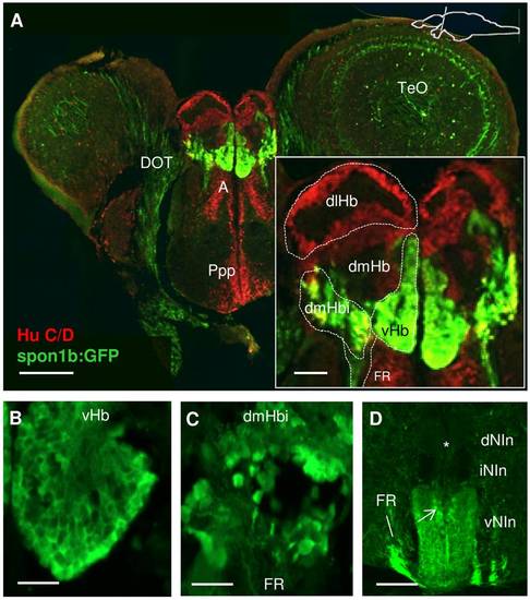
Expression of spon1b and Hu C/D in the habenula (Hb) complex. A. Coronal section through the Hb showing spon1b:GFP immunoreactivity in green and the pan-neuronal marker Hu C/D in red. Note spon1b negative areas at this level: Anterior nucleus of thalamus (A), posterior preoptic areas (ppp). Inset to (A): schematic subdivisions of the Hb in spon1b-positive ventral nucleus (vHb), inferior nucleus of dorsomedial zone (dmHbi), and spon1b-negative dorsolateral (dlHb) and dorsomedial (dmHb) zones. B-C. High magnification images of the small densely packed cells in vHb (B) and the larger sparse cells in dmHbi (C). D. Coronal view of the interpeduncular nucleus (NIn) showing spon1b-positive terminal projections to the ventral area (vNIn), but not the dorsal and intermediate Nin (dNIn, iNIn) (asterisk). Note bypassing fibers from FR circumventing NIn on its way to the SR, and spon1b-positive cells at the core of Nin (arrow). Scale bars: A: 200 μm, inset: 50 μm; B-C: 25 μm; D: 100 μm. |
| Gene: | |
|---|---|
| Fish: | |
| Anatomical Terms: | |
| Stage: | Adult |