Fig. 3
|
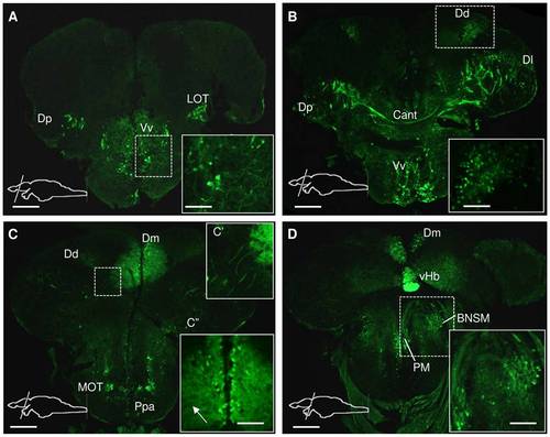
Spon1b expression in the telencephalon of adult zebrafish. Immunostaining for GFP in Tg(spon1b:GFP), coronal sections from rostral (A) to caudal (D) end. A. Spon1b expression in the central nucleus of ventral Tel (Vv), and terminal projections of the lateral olfactory tract (LOT), medial to dorsal nucleus of dorsal Tel (Dp). Inset: thick projections of spon1b-positive cells in Vv. B. Spon1b expression in Vv and the lateral (Dl), posterior (Dp) and dorsal (Dd) nuclei of the dosal Tel. Inset: small positive cells in Dd surrounding sulcus ypsiloniformis. Cant: anterior commissure. C. Robust spon1b expression in the medial nucleus of the dorsal Tel (Dm) and in the medial olfactory tract (MOT). Inset C′: Thick and long projections, originating from LOT area. Inset C′′: medially located spon1b positive cells in Dm with thin and dense projections extending laterally throughout the nucleus (arrow). Ppa: parvocellular preoptic area D. Spon1b expression in the preoptic area. Inset: magnocellular nucleus (PM) and bed nucleus of stria medullaris (BNSM). Strong spon1b expression in the ventral habenula (vHb) (only the right vHb is visible). Scale bars: A-D: 200 Μm. Insets: 50 μm. |

