
Fig. 4
- ID
- ZDB-FIG-120720-15
- Publication
- Harden et al., 2012 - Close association of olfactory placode precursors and cranial neural crest cells does not predestine cell mixing
- Other Figures
- All Figure Page
- Back to All Figure Page
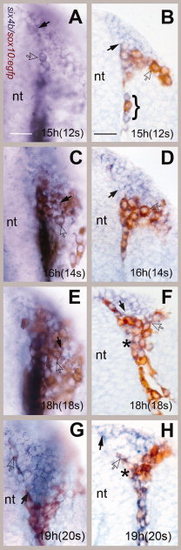
Cranial neural crest cells (CNCCs) move rostrally surrounding the forming olfactory placode (OP). A?H: Whole-mount preparations (A,C,E,G) and cryostat sections (B,D,F,H) of six4b (in situ, blue)/ anti- green fluorescent protein (GFP; immunocytochemistry, brown) double-labeled embryos. All images are dorsal views, rostral to top of the page. A,B: At 12s (somite stage), the neural crest cells were first seen meeting the posterior edge of the OP with some mixing of CNCCs and OP cells (B, bracket). C,D: At 14s, the CNCCs cells began to aggregate at the posterior border of the OP. E,F: At 18s, the neural crest cells surrounded the OP. G,H: The border of the OP was refined at 20s. six4b-expressing cells (black arrows) were observed at the edge of the forming OP. E,F: Some cells (asterisks) appeared to be double labeled. nt, neural tube. Scale bars A (for A?G) and B (for B?H) = 30 μm. Five whole-mount and sectioned embryos were examined at high magnification per time point. |