
Fig. 1
- ID
- ZDB-FIG-120525-25
- Publication
- Gao et al., 2012 - Dcc Regulates Asymmetric Outgrowth of Forebrain Neurons in Zebrafish
- Other Figures
- All Figure Page
- Back to All Figure Page
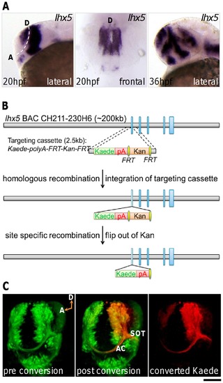
ADt neurons project axons ventrally. In this and subsequent figures, the probes used for whole-mount in situ hybridization are listed in the upper right corner of each panel. Developmental stages are indicated in the lower left corners. Lateral, animals mounted in lateral view, rostral to the left; Frontal, animals mounted in frontal view, dorsal to the top. (A) lhx5 is expressed in the anterior dorsal region of the telencephalon. Dashed line marks the telencephalon-diencephalon border. D: dorsal; A: anterior ventral. Scale bar: 100 μm for lateral view; 60 μm for frontal view. (B) BAC modification via recombination methods. Vertical blue bars represent exons of the lhx5 gene. The Kaede expression cassette replaced the first exon of lhx5 gene. pA: polyadenylation signal sequence; Kan: kanamycin resistant marker; FRT, flippase recognition target. (C) Photo-conversion of Kaede in Tg(lhx5BAC:Kaede) transgenic embryos demonstrates ADt neurons project axons ventrally into the AC and SOT. A live Tg(lhx5BAC:Kaede) transgenic animal was mounted in tilted frontal view to reveal the AC and SOT simultaneously. The region of the left telencephalon was selected for photoconversion. D: dorsal; A: anterior ventral; AC: anterior commissure; SOT: supraoptic tract. Scale bar: 60 μm. |
| Genes: | |
|---|---|
| Fish: | |
| Anatomical Terms: | |
| Stage Range: | 26+ somites to Prim-25 |