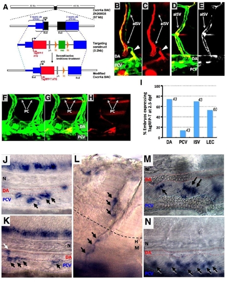
Fig. S2
|
(related to Figure 2). Determination of cxcr4a expression pattern of using recombineered Cxcr4a BAC constructs. (A) Construction of a targeted BAC construct driving TagRFP-T expression from cxcr4a promoter-enhancer sequences. (B-E) Confocal images of Tg(fli1a:EGFP)y1 embryo (green) at 35 hpf (B and C) and 48 hpf (D and E) injected with a recombineered BAC clone with TagRFP-T driven by the cxcr4a promoter (red). TagRFP-T is detected in ISV, DA, and a subset of cells in PCV from which lymphatic progenitor emerges (arrowhead in B and C). (B and D) Merged image. (C and E) Red channel only. (F-H) Confocal images of 2 dpf Tg(fli1a:EGFP)y1 embryo (green) injected with a recombineered BAC clone with TagRFP-T driven by the cxcr4a promoter (red). TagRFP-T is detected in lymphatic progentor sprouting from PCV and PC. (F) Green channel only. (G) Merged image. (H) Red channel only. (I) Quantification of TagRFP-T expression domains in modified Cxcr4a BAC-injected Tg(fli1a:EGFP)y1 embryo at 2.5 dpf. Numbers above the bars show the number of TagRFP-T expressing embryos counted. (J-N) WISH images showing cxcr4b expression in various stages of Zebrafish. Cxcr4b is detected in a subset of PCV cells at 32 hpf (black arrows; J) and at 36 hpf (black arrows; K), in a sprouting lymphatic progenitor (white arrow in K), in dorsoventrally migrating lymphatic cells at 65 hpf (black arrows; L), and in lymphatic cells forming the TD at 3 dpf (black arrows; M) and 4 dpf (black arrows; N). |
Reprinted from Developmental Cell, 22(4), Cha, Y.R., Fujita, M., Butler, M., Isogai, S., Kochhan, E., Siekmann, A.F., and Weinstein, B.M., Chemokine Signaling Directs Trunk Lymphatic Network Formation along the Preexisting Blood Vasculature, 824-836, Copyright (2012) with permission from Elsevier. Full text @ Dev. Cell

