
Fig. 2
- ID
- ZDB-FIG-120424-14
- Publication
- Dente et al., 2011 - Cloning and developmental expression of zebrafish pdzrn3
- Other Figures
- All Figure Page
- Back to All Figure Page
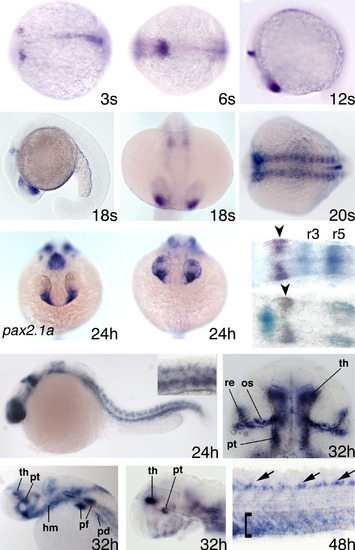
Expression pattern of pdzrn3. (A,B) Three-somite (A) and six-somite (B) stage: dorsal views, anterior to the left. (C-E) twelve-somite (C) and eighteen-somite (D) stage: lateral views, anterior to the left. (E) Eighteen-somite stage: dorsal view, anterior to the bottom. (F) Twenty-somite stage embryo stained for prolonged time: dorsal view, anterior to the left. (G,H) Comparison between the anterior expression of pax2.1a (G) and pdzrn3 (H) at 24-hpf stage: frontal views, dorsal to the top. (I,J) Comparison between the rhomboencephalic expressions of pdzrn3 (I, J; purple staining indicated by arrowheads), krox20 (I, turquoise staining) and pax2.1a (J, turquoise staining); dorsal view, anterior to the right. (K) 24-hpf stage embryo stained for prolonged time: lateral view, anterior to the left. The inset in the top right corner shows a magnification of somite expression in the trunk. (L) Head expression in 32 hpf embryo: ventral view anterior to the top; os, optic stalk; pt, posterior tuberculum; re, retina; th, thalamus. (M,N) 32 hpf stage: lateral view, anterior to the left; hm, head mesenchyme; pd, pronephric duct; pf, pectoral fin. To analyze internal brain expression sites the eyes of the embryo shown in N were removed. (O) Trunk and tail expression at 48 hpf: dorsal to the top, anterior to the left. Arrows point to the motor neurons of the spinal cord. The bracket delimits pdzrn3 expression in the ventral region of somites. |