
FIGURE
Fig. S5
Fig. S5
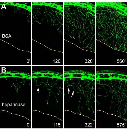
Peripheral Axons Avoid Heparinase III-Injected Areas, Related to Figure 6 Representative still images from time-lapse confocal series (Movie S5 and Movie S6) showing peripheral axon outgrowth in BSA- (A) and heparinase III-injected (B) embryos. Yellow dotted lines indicate ventral edge of the embryos. Arrows indicate axons that appear to be encountering the heparinase-injected region and fail to grow ventrally, instead branching or turning. |
Expression Data
Expression Detail
Antibody Labeling
Phenotype Data
Phenotype Detail
Acknowledgments
This image is the copyrighted work of the attributed author or publisher, and
ZFIN has permission only to display this image to its users.
Additional permissions should be obtained from the applicable author or publisher of the image.
Full text @ Curr. Biol.