
Fig. 5
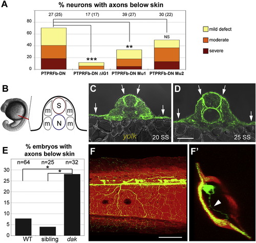
HSPG Binding to LAR Proteins Is Likely Important for Skin Innervation (A) Disrupting the first Ig domain significantly reduced skin innervation defects caused by expressing PTPRFb-DN. Deleting Ig1 or mutating a proteoglycan-binding-site (Mu1) in PTPRFb-DN significantly reduced skin innervation defects (11.8% and 33.3%, respectively). The number of neurons is listed at the top of each bar; the number of fish is listed in parenthesis.(B) Diagram of <20 SS zebrafish embryo cross-section. Red line indicates position of cross-section. S, spinal cord; N, notochord; m, muscle. C and D) Cross-sections of anti-HS staining at 20 and 25 SS. HSPGs were enriched in basement membranes around the spinal cord, notochord, and skin. Arrows indicate high levels of anti-HS staining in the skin. Scale bar represents 50 �m. (E) Frequency of embryos with peripheral axons below the skin in a defined region of the trunk (see Figure 2). Peripheral axons in this region were found below the skin in 28.1% of dackel homozygous mutant embryos, which was significantly higher than in their nonmutant siblings (4.0%). (F) Lateral view of 3 dpf dackel mutant larva harboring krt4:dsRed and sensory:GFP transgenes. (F2) Ninety degree rotation of image in (F). Arrowhead indicates axon innervating internal tissue. All data analyzed with two-sided Fisher′s exact test and computed with R. ***p < 0.001; **p < 0.01; *p < 0.05. Scale bar represents 100 μm. See also Figure S4. |
| Fish: | |
|---|---|
| Observed In: | |
| Stage: | Long-pec |