
Fig. 3
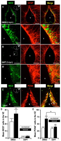
Notch intracellular domain immunoreactivity after telencephalic injury. (A,B) Immunodetection of the Notch intracellular domain (NICD) in coronal brain sections (dorsal up) at 1 dpl. Higher magnifications of panels A and B are shown in panels A′ and B′, respectively. (C) Immunodetection of a proliferation marker (PCNA) and the NICD in a coronal brain section (dorsal up) at 1 dpl. Arrowheads indicate NICD and PCNA double-positive cells in the telencephalic VZ. V, telencephalic ventricle. (D,E) Histograms showing the counts of NICD-positive cells (D) and PCNA-positive cells (E) in the telencephalic VZ. Notice the accumulation of NICD in proliferating cells in the telencephalic VZ induced in the injured hemisphere, compared with the uninjured one (A,C,D). This accumulation was prevented by treatment with the Notch inhibitor DAPT (B,D,E). Student?s t-test was used to determine significant differences in expression. Error bars represent s.e.m. **P<0.01, ***P<0.001. Scale bars: 50 μm (A,B,C); 10 μm (A′,B′). |