
Fig. 2
- ID
- ZDB-FIG-120315-46
- Publication
- Simmons et al., 2012 - Mutation of pescadillo Disrupts Oligodendrocyte Formation in Zebrafish
- Other Figures
- All Figure Page
- Back to All Figure Page
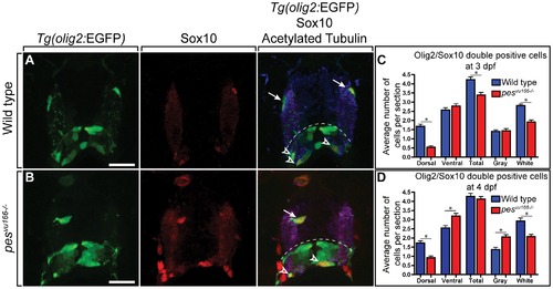
Distribution of oligodendrocyte lineage cells in wild-type and pesvu166 mutant larvae. Transverse sections through spinal cords of 4 dpf wild-type (A) and pesvu166 -/- (B) larvae carrying the Tg(olig2:EGFP) reporter and processed for Sox10 immunohistochemistry. (A) Wild type spinal cord section showing normal number and distribution of dorsal (arrows) and ventral (arrowheads) olig2+, Sox10+ oligodendrocytes. Dashed line marks position of pMN precursor domain. Anti-Acetylated Tubulin staining (blue) marks axon-rich white matter region of spinal cord. Dorsal olig2+, Sox10+ oligodendrocytes occupied white matter. (B) pesvu166 -/- spinal cord section. Dorsal olig2+, Sox10+ oligodendrocyte (arrow) was located in gray matter region, medial to the white matter region. (C,D) Quantification of oligodendrocytes in the dorsal, ventral, gray matter, and white matter spinal cord regions at 3 and 4 dpf. Ten sections from ten larvae of each genotype were counted. Scale bar represents 30 μm. Asterisk (*) indicates pj0.05 by Student′s T-test. |
| Genes: | |
|---|---|
| Antibody: | |
| Fish: | |
| Anatomical Term: | |
| Stage: | Day 4 |
| Fish: | |
|---|---|
| Observed In: | |
| Stage Range: | Protruding-mouth to Day 4 |