
Fig. 4
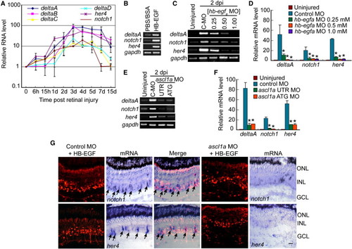
HB-EGF and Ascl1a Regulate Notch-Signaling Component Genes in the Injured Retina (A) Real-time PCR shows injury-dependent induction of Notch-signaling components. (B) RT-PCR shows that HB-EGF activates Notch-signaling component genes in the uninjured retina. (C) RT-PCR shows that MO-mediated HB-EGFa knockdown suppresses injury-dependent induction of Notch-signaling component genes. (D) Real-time PCR quantification of (C). *p < 0.01. (E) RT-PCR shows that Ascl1a knockdown suppresses injury-dependent induction of Notch-signaling component genes. (F) Real-time PCR quantification of (E). *p < 0.01. (G) Ascl1a knockdown suppresses HB-EGF-dependent induction of notch1 and her4 mRNAs. Arrows identify notch1 and her4 mRNA expression in control MO-treated cells. Scale bar, 50 μm. Error bars represent SD. ONL, outer nuclear layer; INL, inner nuclear layer; GCL, ganglion cell layer. See also Figures S4 and S5. |
| Genes: | |
|---|---|
| Fish: | |
| Condition: | |
| Knockdown Reagents: | |
| Anatomical Term: | |
| Stage: | Adult |
Reprinted from Developmental Cell, 22(2), Wan, J., Ramachandran, R., and Goldman, D., HB-EGF Is Necessary and Sufficient for M�ller Glia Dedifferentiation and Retina Regeneration, 334-347, Copyright (2012) with permission from Elsevier. Full text @ Dev. Cell