
Fig. 6
- ID
- ZDB-FIG-120214-4
- Publication
- Malicki et al., 1996 - Mutations affecting development of the zebrafish retina
- Other Figures
- All Figure Page
- Back to All Figure Page
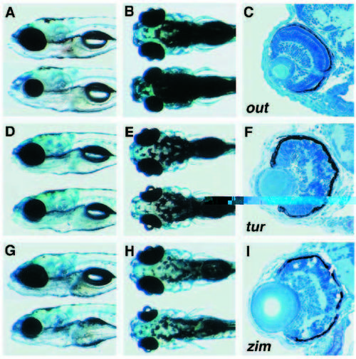
Examples of mutant phenotypes from the categories of Growth retardation and Nonspecific retinal degeneration. Mutant individuals (lower) are shown next to their wild-type siblings (upper). (A,B,C) out of sight (out)m233. (D,E,F) turbulent (tub)m125. (G,H,I) zimny (zny)m419. (A,B) The eye of outm233 is substantially reduced at 3 dpf. (C) All retinal laminae are present and cell death in excess of wild-type levels is not observed in outm233 at this stage. At 5 dpf mutants tubm125 (D,E) and znym419(G,H) are characterized by a reduced eye size and somewhat abnormal brain shape. Cell death is extensive in both tubm125 (F) and znym419(I) retinae at 3 dpf. Cell corpses appear as small, round, intensely staining particles (F). Retinal patterning defects are inconsistent in this group of mutants. A, D and G show lateral views; B, E and H dorsal views. The dorsal side is oriented up in panels showing lateral views or sections. In panels showing head phenotypes anterior is left. All sections are transverse. |
| Fish: | |
|---|---|
| Observed In: | |
| Stage Range: | Protruding-mouth to Day 5 |