
Fig. 5
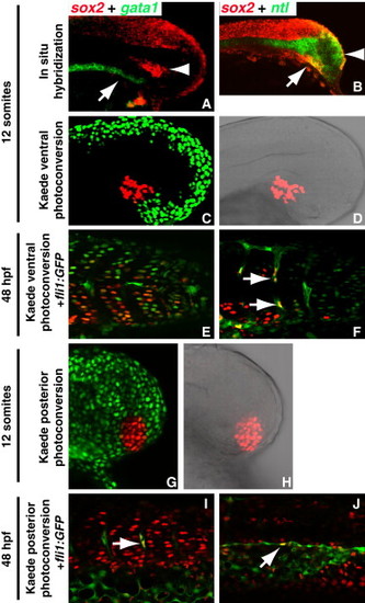
Mesodermal Progenitors of the Tail Bud Normally Contribute to Posterior Endothelial Tissue (A) Double fluorescent in situ staining of gata1 (green, arrow) and sox2 (red, arrowhead) indicates that the sox2 positive cells of the tail bud are distinct from the gata1 expressing intermediate cell mass. (B) Double fluorescent in situ staining of ntl (green) and sox2 (red) indicates that sox2 positive cells coexpress ntl in both the ventral domain (arrow) and in the SZ (arrowhead). (C?J) fli1:gfp transgenic embryos were injected with nuclear localized kaede mRNA (C and D). A small population of cells were photoconverted from green to red in the region of the ventral sox2 expressing cells, (G and H) and in the SZ region of sox2 expressing cells. (E, F, I, and J) In both cases, cells gave rise to both somites and vascular endothelium (arrows indicate red nucleus in a cell with cytoplasmic GFP expression from the fli1:gfp). |
Reprinted from Developmental Cell, 22(1), Martin, B.L., and Kimelman, D., Canonical Wnt Signaling Dynamically Controls Multiple Stem Cell Fate Decisions during Vertebrate Body Formation, 223-232, Copyright (2012) with permission from Elsevier. Full text @ Dev. Cell