Fig. 3
- ID
- ZDB-FIG-120201-28
- Publication
- Chen et al., 2011 - Role of zebrafish lbx2 in embryonic lateral line development
- Other Figures
- All Figure Page
- Back to All Figure Page
|
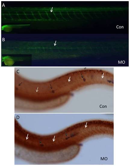
Disassociation of the PLL primodium and PLL nerve in lbx2 morphants. (A, B) Labeling of the PLL nerve using an anti-acetylated α-tubulin antibody, indicating that the PLL nerve grew correctly in both control embryos (A) and lbx2 morphants (B). (C, D) Immunohistochemical detection of the PLL nerve using an anti-acetylated alpha tubulin antibody to label PLL neuromasts and in situ hybridization using a cldnb antisense probe to label the primordia, revealing disassociation of the primodium and nerve in lbx2 morphants (D), but not in control embryos (C). White arrows indicate the PLL nerve, black arrows show the deposited neuromasts and the black asterisk indicates the stalled primordium. Embryos used in the assay were at 48 hpf stage. |

