Fig. S1
- ID
- ZDB-FIG-120201-24
- Publication
- Chen et al., 2011 - Role of zebrafish lbx2 in embryonic lateral line development
- Other Figures
- All Figure Page
- Back to All Figure Page
|
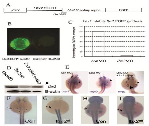
Efficiency of the lbx2 morpholino. (A) A test lbx2-EGFP construct was created containing 60 bp of the 5′ UTR and the first 66 amino acid coding sequence of lbx2 cDNA fused to the N-terminus of EGFP, driven by the CMV promoter. The sequence of lbx2 MO is complementary to the 1–24 bp region of zebrafish lbx2 cDNA. (B) Live embryos at the 50% epiboly stage. Embryos co-injected with 25 ng lbx2-EGFP DNA and 5 ng control MO expressed green fluorescent fusion protein (left), which was inhibited by co-injection of 2 ng lbx2 MO (right). (C) Translation of lbx2-EGFP in live embryos was inhibited by co-injection of lbx2 MOs. (D) The lbx2 protein level in lbx2-MO-injected embryos was drastically lower than control MO-injected embryos at 30 hpf (E). Absence of MyoD expression in the pectoral fin bud of lbx2 morphants at 48 hpf, which could be rescued by co-injection of lbx2 mRNA. Arrowhead indicates MyoD expression in the pectoral fin bud area. (F–I) Injection of lbx2eh- mRNA dramatically inhibited MyoD expression in pectoral fin muscle precursors at 30 hpf (G) and 36 hpf (I), compared to gfp mRNA-injected control embryos (F, H). |

