Fig. 3
- ID
- ZDB-FIG-111020-12
- Publication
- de Borsetti et al., 2011 - Light and melatonin schedule neuronal differentiation in the habenular nuclei
- Other Figures
- All Figure Page
- Back to All Figure Page
|
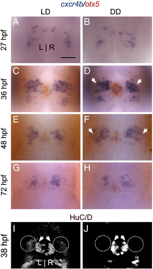
Habenular progenitor cells accumulate in an undifferentiated state in constant darkness. (A?B) A similar number of cxcr4b-positive habenular progenitor cells are specified at 27 hpf in LD and DD. (C?F) However, at 36 and 48 hpf, many more progenitor cells accumulate in DD conditions than in LD. (G?H) By 72 hpf, only a few progenitor cells are detected in LD or DD. (I?J) At 38 hpf, fewer HuC/D-positive post-mitotic precursor cells are detected in the habenular nuclei (white ovals), consistent with the retention of habenular cells in a progenitor state. HuC/D-positive projection neurons in the pineal complex (white arrowheads), meanwhile, are unaffected by DD conditions (average of 22 cells in LD versus 21 cells in DD, p > 0.42 in two-tailed t-test). Image in J is intentionally overexposed to confirm the absence of HuC/D signal in the habenular nuclei. All views are dorsal. Scale bar = 50 μm. |
Reprinted from Developmental Biology, 358(1), de Borsetti, N.H., Dean, B.J., Bain, E.J., Clanton, J.A., Taylor, R.W., and Gamse, J.T., Light and melatonin schedule neuronal differentiation in the habenular nuclei, 251-61, Copyright (2011) with permission from Elsevier. Full text @ Dev. Biol.

