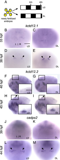
Fig. 2
- ID
- ZDB-FIG-111020-11
- Publication
- de Borsetti et al., 2011 - Light and melatonin schedule neuronal differentiation in the habenular nuclei
- Other Figures
- All Figure Page
- Back to All Figure Page
|
Reversal of the photoperiod phase only slightly advances the timing of gene expression in the habenular nuclei. (A) Zebrafish embryos were raised in 14/10 light/dark (LD) or 10/14 dark/light (DL) conditions beginning at 5 min post-fertilization. (B?E) Expression of kctd12.1 is absent at 33 hpf in the habenular nuclei and initiates at 38 hours post-fertilization (hpf) in LD (black arrows). Expression is higher in DL than in LD embryos. (F?M) Expression of kctd12.2 and cadps2 initiates at the same time in LD and DL conditions. Insets in F?I are magnified views of the left habenula. All views are dorsal except for lateral views in F?I. Scale bar = 50 μm except for insets in F?I (25 μm). |
Reprinted from Developmental Biology, 358(1), de Borsetti, N.H., Dean, B.J., Bain, E.J., Clanton, J.A., Taylor, R.W., and Gamse, J.T., Light and melatonin schedule neuronal differentiation in the habenular nuclei, 251-61, Copyright (2011) with permission from Elsevier. Full text @ Dev. Biol.

