|
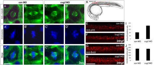
Loss of cug2 function leads to defective mitosis. A-D′′. Anti-α-tubulin immunostaining of the control MO- (A-A′′, B-B′′) and cug2 MO-injected embryos (C-C′′, D-D′′) at the 3-somite stage. cug2 morphants display defective spindle formation and misaligned chromosomes at the metaphase plate (C′, D′). Scale bar = 10 μm. E-H. Lateral views of the spinal cord at 24 hpf. E, F. Anti-phospho-histone H3 immunostaining of control and cug2 morphants. G, H. Anti-BrdU immunostaining of control and cug2 morphants. Scale bar = 100 μm. I, J. Quantification of pH3- (I) and BrdU-positive cells (J) in control and cug2 morphants at 24 hpf (n = 10). The cells were counted from the trunk region, in an area of spinal cord schematically shown (red box) in K.
|