
Fig. 6
- ID
- ZDB-FIG-110706-20
- Publication
- Budi et al., 2011 - Post-embryonic nerve-associated precursors to adult pigment cells: genetic requirements and dynamics of morphogenesis and differentiation
- Other Figures
- All Figure Page
- Back to All Figure Page
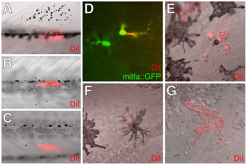
DiI-labeling showed extra-hypodermal contributions to metamorphic melanophores and iridophores. (A?C) DiI labeled tissues imaged immediately after injection into the base of the dorsal fin (A), the vicinity of the horizontal myoseptum and lateral line nerve (B), and the inner myotome (C). Each site yielded hypodermal DiI+; mitfa::GFP+ cells or DiI+ melanophores (12 of 30 larvae, 3 of 30 larvae, 15 of 87 larvae, respectively). (D?F) DiI+ cells that expressed either mitfa::GFP (D) or contained melanin (E,F) found within the lateral hypodermis 4 d following injection into the base of the dorsal fin (D, F) or the inner myotome (E). (G) DiI-labeling was observed for additional cells including iridophores. Although the frequencies with which DiI labeled pigment cells were found differed between injection sites, each site gave rise to DiI+ iridophores at a frequency indistinguishable from that of DiI+ mitfa::GFP+ cells (χ2 = 0.6, d.f. = 1, P = 0.4). We did not observe DiI-labeled xanthophores. |