
Fig. 1
- ID
- ZDB-FIG-110706-16
- Publication
- Budi et al., 2011 - Post-embryonic nerve-associated precursors to adult pigment cells: genetic requirements and dynamics of morphogenesis and differentiation
- Other Figures
- All Figure Page
- Back to All Figure Page
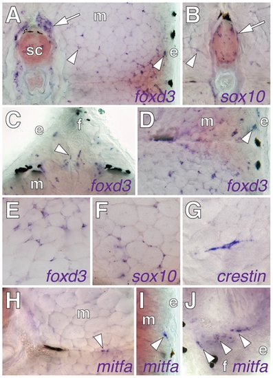
Post-embryonic expression of embryonic neural crest and glial markers. Shown are in situ hybridizations performed on transverse sections of 7?9 SSL larvae. (A) foxd3 transcript was detected in dorsal root ganglia (arrow) and scattered cells (e.g., arrowheads) within the myotome (m) and near the epidermis (e). (B) sox10 expression by cells adjacent to the neural tube (arrow) and within the myotome (arrowhead). (C) foxd3+ cells (e.g., arrowheads) at the base of the dorsal fin (f). (D) foxd3+ cells within the myotomes and near the epidermis. (E?G) foxd3+, sox10+, and crestin+ cells within the myotomes. (H?J) Cells expressed mitfa (arrowheads), within the horizontal myoseptum (H), at the surface of the myotome (I), and at the base of anal fin (J) (see text for details). |