
Fig. 4
- ID
- ZDB-FIG-110422-11
- Publication
- Kawahara et al., 2011 - Drug screening in a zebrafish model of Duchenne muscular dystrophy
- Other Figures
- All Figure Page
- Back to All Figure Page
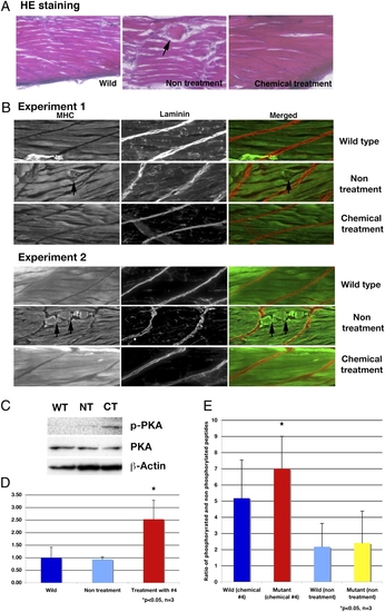
Analysis of skeletal muscle of dystrophin-null fish that survive 30 d after treatment with chemical 4, aminophylline. (A) H&E staining of WT, nontreated, and chemical 4-treated fish. Nontreated dystrophin-null fish have disorganized structure (arrow) in skeletal muscle. The dystrophin-null fish with treated aminophylline have normal structure in skeletal muscle. (B) Immunostaining of WT, nontreated, and chemical 4-treated fish with anti-myosin heavy chain and anti-laminin antibody. Nontreated dystrophin-null fish have broken and disturbed structure (arrows) of skeletal muscle fibers. The treated dystrophin-null fish with aminophylline have normal structure in skeletal muscle. (C) Immunoblot of phosphorylated PKA and PKA at 30 dpf in surviving fish. WT, wild type; NT, nontreatment of dystrophin-null fish; CT, chemical 4-treated dystrophin-null fish. (D) Ratio of phosphorylated PKA and PKA in 30 dpf surviving fish (n = 3). Blue, WT; light blue, nontreatment of dystrophin-null fish; red, chemical 4-treated dystrophin-null fish. *P < 0.05 (vs. WT and nontreatment groups). Error bars indicate SDs. (E) Assay of cAMP-dependent PK activity (n = 3). Bars show the ratio of phosphorylated peptides and nonphosphorylated peptide. Blue, WT fish treated with chemical 4 (2.5 μg/mL) for 30 d. Red, affected sapje fish treated with chemical 4 (2.5 μg/mL). Light blue, nontreated WT fish. Yellow, nontreated affected sapje fish. *P < 0.05 (vs. nontreated WT and mutant fish). Error bars indicate SDs. |
| Fish: | |
|---|---|
| Condition: | |
| Observed In: | |
| Stage: | Days 30-44 |