
Fig. 3
- ID
- ZDB-FIG-110322-70
- Publication
- Burroughs-Garcia et al., 2011 - Evolutionarily conserved function of Gbx2 in anterior hindbrain development
- Other Figures
- All Figure Page
- Back to All Figure Page
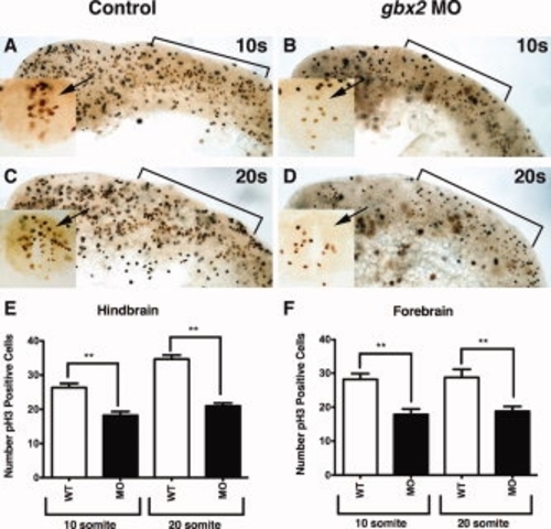
Reduced proliferation in gbx2 morphant embryos. A?F: Embryos were labeled with the anti-pH3 antibody to label cells undergoing mitosis. A,B: Lateral views of uninjected and embryo injected with 8 ng of gbx2-MO illustrates that the decrease in mitotic figures occurs at 10-somites. Inset (A?D): Transverse section demonstrates mitotic figures in the neural tube of the r2 domain (arrow). C,D: Lateral views at 20-somite stage shows that a decrease in pH3-positive cells persists in gbx2 morphant embryos. E: Number of pH3-positive cells in the r2 domain of the anterior hindbrain as indicated in the insets. F: Number of pH3-positive cells in the forebrain. Brackets, anterior hindbrain; MO, morpholino; s, somite stage. |
| Fish: | |
|---|---|
| Knockdown Reagent: | |
| Observed In: | |
| Stage Range: | 10-13 somites to 20-25 somites |