
|
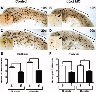
Fig. 3
Reduced proliferation in gbx2 morphant embryos. A?F: Embryos were labeled with the anti-pH3 antibody to label cells undergoing mitosis. A,B: Lateral views of uninjected and embryo injected with 8 ng of gbx2-MO illustrates that the decrease in mitotic figures occurs at 10-somites. Inset (A?D): Transverse section demonstrates mitotic figures in the neural tube of the r2 domain (arrow). C,D: Lateral views at 20-somite stage shows that a decrease in pH3-positive cells persists in gbx2 morphant embryos. E: Number of pH3-positive cells in the r2 domain of the anterior hindbrain as indicated in the insets. F: Number of pH3-positive cells in the forebrain. Brackets, anterior hindbrain; MO, morpholino; s, somite stage.