
Fig. 6
- ID
- ZDB-FIG-110322-48
- Publication
- Veth et al., 2011 - Mutations in Zebrafish lrp2 Result in Adult-Onset Ocular Pathogenesis That Models Myopia and Other Risk Factors for Glaucoma
- Other Figures
- All Figure Page
- Back to All Figure Page
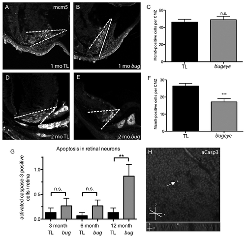
Retinal proliferation and apoptosis. A?F Mcm5 expression in 1 (A?C) and 2-month (D?F) cryosections. Dashed white lines denote proliferative ciliary marginal zone (CMZ) of the retina; asterisks indicate autofluorescent blood vessels. G Quantitation of apoptotic cells identified on whole retina flat-mounts by activated caspase-3 (aCasp3) immunofluorescence. H Confocal images of aCasp3-positive cell in 1 year old bugeye mutant. Upper shows compressed z-stacks, lower shows 90°rotation to reveal z location of positive cell (arrow). The flat mounted retinas were orientated with retinal ganglion cell layer up. n = 15 eyes for each condition; ***p<0.001, **p<0.01, n.s., not significant (t-test). |
| Fish: | |
|---|---|
| Observed In: | |
| Stage Range: | Days 45-89 to Adult |