
FIGURE
Fig. 3
- ID
- ZDB-FIG-110121-1
- Publication
- Seiler et al., 2011 - Transgenic labeling of the zebrafish pronephric duct and tubules using a promoter from the enpep gene
- Other Figures
- All Figure Page
- Back to All Figure Page
Fig. 3
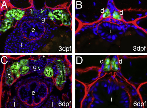
GFP expression in kidney tubules and ducts: Cross section of 3 dpf (A, B) and 6 dpf (C, D); Tg(enpep:GFP) larva stained with anti-GPF (green) and anti-Laminin (red) antibodies. GFP expression can be detected in both pronephric tubules (?t?, A, C) and ducts (?d?, B, D) as well as in podocyte like cells (arrowheads) of the glomerulus (?g? A, C). At 6 dpf several loops of the tubules are present (C). No expression is present in the intestine (i), esophagus (e) or liver (l). |
Expression Data
| Gene: | |
|---|---|
| Fish: | |
| Anatomical Terms: | |
| Stage Range: | Protruding-mouth to Day 6 |
Expression Detail
Antibody Labeling
Phenotype Data
Phenotype Detail
Acknowledgments
This image is the copyrighted work of the attributed author or publisher, and
ZFIN has permission only to display this image to its users.
Additional permissions should be obtained from the applicable author or publisher of the image.
Reprinted from Gene expression patterns : GEP, 11(1-2), Seiler, C., and Pack, M., Transgenic labeling of the zebrafish pronephric duct and tubules using a promoter from the enpep gene, 118-121, Copyright (2011) with permission from Elsevier. Full text @ Gene Expr. Patterns