|
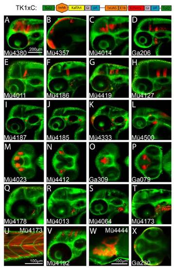
Transgenic zebrafish KalTA4 enhancer trap strains. A schematic representation of the trapping construct TK1xC is shown at the top of the figure. (A?X) Confocal microscopy images of Bodipy ceramide-counterstained (green) embryos from isolated KalTA4 enhancer trap lines. Embryos were screened for tissue-specific mCherry fluorescence (red) between 24 and 36 hpf; images were recorded at approximately 50 hpf using a Zeiss LSM510 and a 20x objective. (A?L, Q?T, and U?W) lateral views (W 6dpf heart), (M?P) ventral views and (X) dorsal view. For detailed description of expression patterns see the database at: http://www.helmholtz-muenchen.de/en/idg/groups/neuroimaging/lines_distel/.
|