
Fig. S1
- ID
- ZDB-FIG-101115-28
- Publication
- Qin et al., 2009 - Genetic evidence for shared mechanisms of epimorphic regeneration in zebrafish
- Other Figures
- All Figure Page
- Back to All Figure Page
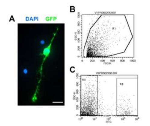
Isolation of GFP+ Müller glia. (A) Dissociated GFP+ Müller glial cell (green). Counterstained with DAPI (blue). (B and C) Flow cytometry scatter plots. forward scatter-height (FSC-H); side scatter-height (SSC-H). Dissociated cells from adult Tg(gfap:GFP)mi2002 zebrafish retinas were gated by forward and side scatters (B), and GFP+ Müller glia were isolated based on fluorescence in the FITC channel (R5) (C). Our yield of dissociated retinal cells from adult zebrafish (5- to 6-month old) was ~2.5 x 10⁵ cells/retina, of which ~9% were GFP+ Müller glia. With flow cytometry, we could recover ~2.1 x 10⁴ Müller glia/retina, representing an efficiency of ~84%. (Scale bar: 10 μm.) |