
Fig. 4
- ID
- ZDB-FIG-101111-44
- Publication
- Prykhozhij, 2010 - In the Absence of Sonic Hedgehog, p53 Induces Apoptosis and Inhibits Retinal Cell Proliferation, Cell-Cycle Exit and Differentiation in Zebrafish
- Other Figures
- All Figure Page
- Back to All Figure Page
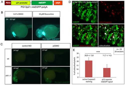
(A) p53 reporter PG13p21::nlsEGFP-polyA was constructed using Tol2kit as described in Materials and methods. It consists of the p21 promoter fused with p53-binding enhancer sequence PG13, nlsEGFP coding sequence and a polyadenylation signal (polyA). (B) Wild-type embryos carrying the p53 reporter transgene were incubated for 6 hours at 24 hpf with either 50 μM Roscovitine or 0.4% DMSO diluted in E3 medium. DMSO-treated embryos did not show any significant expression of the reporter (right), whereas roscovitine-treated ones expressed p53 reporter at a high level (left). All embryos showed green-heart transgenesis marker. The experiment was repeated three times and 20 embryos were used for each treatment. (C) p53 morpholino but not 4-mismatch control morpholino injection suppressed expression of PG13p21::nlsEGFP p53 reporter in shh-/- mutant embryos at 24 hpf. Both injections had no effect on p53 reporter expression in the wild-type embryos. (D) Anti-active Caspase3 staining of cells in the shh-/- mutant retina at 56 hpf expressing p53 reporter transgene PG13p21::nlsEGFP. Most cells positive for active Caspase3 also expressed p53 reporter transgene. Single channel, overlay and co-localisation analysis images are shown. (E) Quantification of co-localisation of active Caspase3 staining and nlsEGFP p53 reporter signal. Co-localised pixels made up 20,9 +/- 3,8% of total active Caspase3 pixels and 11,7 +/ 4,5% of total nlsEGFP pixels. Quantifications were performed on 20 retinal section images from 10 embryos. Asterisks (***) indicate a significant statistical difference between the proportions (t-test, P-value<0,001). |