|
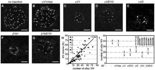
αV and β1b knockdown disrupts physical properties of Kupffer′s vesicle. (A-G) Dorsal views of 6-8 SS embryos; confocal images of Kupffer′s vesicle (KV) cilia were detected by a fluorescent anti-acetylated tubulin antibody. Shown is a 3D rendering of multiple focal planes through the embryo at the level of KV. Total number of embryos used to determine number of cilia per KV and cilia length per morphant: uninjected (n=19); 1.75 ng αV1miss (n=16); 1.25 ng αV1 (n=12); 5 ng αVEI10 (n=9); 1.75 ng αV2 (n=9); 1.0 ng β1b1 (n=8); 5 ng β1bEI10 (n=10). Scale bars: 20 μm. (H) Graphic representation of KV volume versus cilia number per KV. (I) Graphic representation of the number of cilia per KV in control and knockdown embryos. Inset, cilia length in αV and β1b morphants, indicated with their respective panel labels A to G. Data represent mean ± s.e.m.
|