
Fig. 8
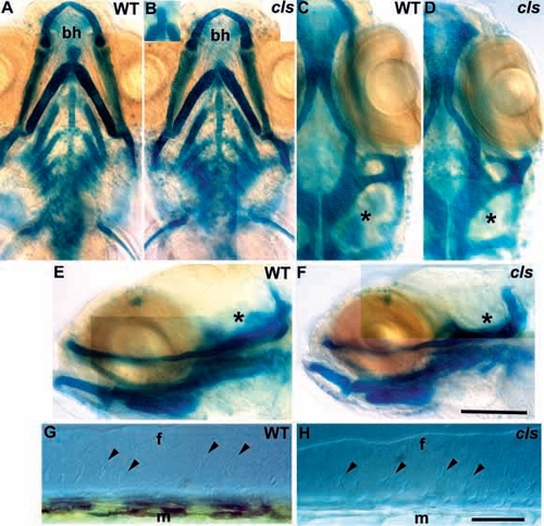
Ectomesenchymal crest derivatives are essentially normal in cls- embryos. Alcian green stained craniofacial skeletons of 5 dpf wild-type (A,C,E) and cls- (B,D,F) larvae are shown in ventral (A,B), dorsal (C,D) and lateral (E,F) views. Mutant craniofacial skeletons are barely distinguishable from wild type, although development is slightly retarded (compare angle of main arch elements in A and B) and the basihyal cartilage is variable (bh in A and B, also inset in B). Ventral (C,D) and lateral (E,F) views show slight differences in the cartilages associated with the developing inner ear (asterisk) which remains small in cls-. Median fin mesenchyme (arrowheads) of the dorsal fin in lateral view with Nomarski optics in 5 dpf wild-type (G) and cls- (H) larvae; the number and position of cells is identical. f, medial fin; m, muscle blocks. Scale bar, 100 μm (A-F) and 50 μm (G,H). |