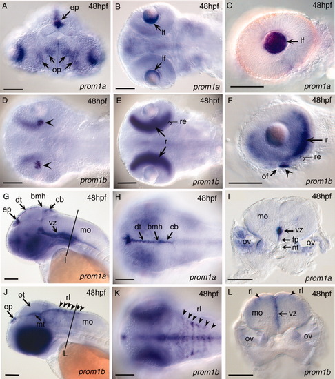
Expression of zebrafish prom1a and prom1b in the sensory organs and CNS of the zebrafish embryo at 48hpf. prom1a (A-C, G-I) and prom1b (D-F, J-L) expression. Sensory organs: prom1a expression in the epiphysis (A, G), olfactory epithelium of the olfactory placodes (A), and lens epithelium of the eye (B, C). prom1b expression in the epipyhysis (J), retinal epithelium (D, F, arrowheads), and marginal layer of the neural retina in the eye (E, F). CNS: G-I: prom1a expression in the tectum and ventricular zones of the hindbrain. J-L: prom1b expression in the midbrain tegmentum and ventricular zones of the hindbrain. A: Anterior frontal view; B, E: dorsal medial optical section; D: ventral view; G, J: lateral view; H, K: dorsal view. I, L: cross-section through the hindbrain neural tube. bmh, boundary between mid- and hindbrain; cb, cerebellum; dt, dorsal optic tectum; ep, epiphysis; fp, floor plate; lf, lens fiber cells; mo, medulla oblongata; mt, midbrain tegmentum; nt, notochord; of, optic fissure; op, olfactory placode; ot, optic tectum; ov, otic vesicle; r, neural retina; re, retinanl epithelium; rl, rhombic lip; vz, ventricular zone. Scale bars = 100 μm.
|

