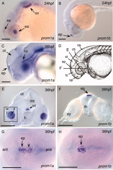
Expression of zebrafish prom1a and prom1b in developing sensory organs and CNS of 24- and 36hpf embryos. A: Lateral view, 24hpf. prom1a expression in the lens anlage, epiphysis, and otic vesicle. B: Lateral view, 24hpf. prom1b expression in the epiphysis (arrowhead). C: Lateral view, 36hpf. prom1a expression in the ventricular zone along the dorsal surface of the midbrain tegmentum and extending into the hindbrain. D: Camera lucida drawing of 35hpf zebrafish embryo (Kimmel et al.,[1995]) showing position of cross-sections shown in E and F. E: At 36hpf, prom1a expression in the ventricular zone of the midbrain tegmentum and lens fiber cells (inset). F: At 36hpf, prom1b expression in the epiphysis. G: Dorsal view, 36hpf. prom1a expression extends from the epiphysis posteriorly along the tectum (arrowhead). H: Dorsal view, 36hpf. prom1b expression is restricted to the epiphysis. ant, anterior; cb, cerebellum; ea, eye anlage; ep, epiphysis; ey, eye; h, hypothalamus; hb, hindbrain; le, lens epithelium; lf, lens fiber cells; mt, midbrain tegmentum; op, olfactory placode; ot, optic tectum; ov, otic vesicle; pos, posterior; tc, telencephalon; th, thalamus; vz, ventricular zone. Scale bars = 100 μm.
|
