
Fig. 2
- ID
- ZDB-FIG-100616-27
- Publication
- Ito et al., 2010 - Characterization of neural stem cells and their progeny in the adult zebrafish optic tectum
- Other Figures
- All Figure Page
- Back to All Figure Page
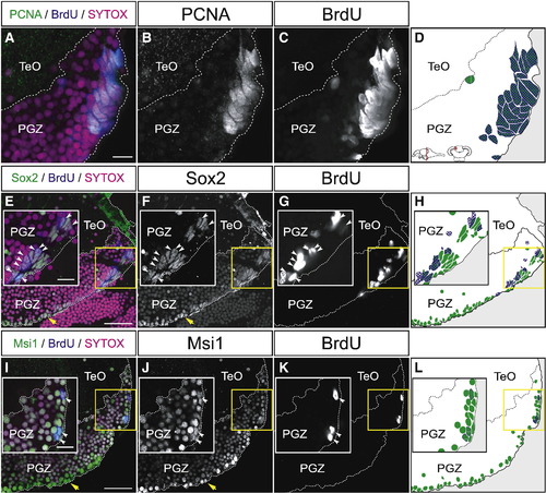
Proliferating cells in the dorsomedial area of the PGZ express neural stem/progenitor cell markers. (A?L) Expression of PCNA (A?D), Sox2 (E?H), and Msi1 (I?L) in the dorsomedial area of the PGZ of the adult zebrafish optic tectum (60 μm transverse sections, single planes, dorsal top). Proliferating cells are labeled with BrdU after 72 h of incubation. Insets in E?L show magnified views of the yellow-boxed areas. (A?D) Most PCNA-positive cells incorporate BrdU after 72 h of BrdU administration. (E?F) The majority of BrdU-positive proliferating cells (insets, arrowheads), and the cells that reside in the ventral edge of the PGZ (yellow arrows) express Sox2. (I?L) A subset of BrdU-positive cells (insets, arrowheads), and the cells that reside in the ventral edge of the PGZ (yellow arrows) express Msi1. CCe, corpus cerebelli; PGZ, periventricular gray zone; TeO, tectum opticum. Scale bars: 10 μm in A, insets of E, I; 30 μm in E, I. |
Reprinted from Developmental Biology, 342(1), Ito, Y., Tanaka, H., Okamoto, H., and Ohshima, T., Characterization of neural stem cells and their progeny in the adult zebrafish optic tectum, 26-38, Copyright (2010) with permission from Elsevier. Full text @ Dev. Biol.