
FIGURE
Fig. 4
Fig. 4
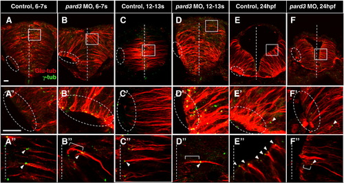
Pard3 is required for apical localization of the centrosome. Sections of control (A?A″, C?C″, E?E″) and pard3 MO-injected (B?B″, D?D″, F?F″) embryos at the early rod (6?7 som, A?B″), late rod (12?13 som, C?D″) and neural tube (24 hpf, E?F″) stages immunolabeled with anti-Glu and anti-γ-tub. Ovals and rectangles in (A?F): areas magnified in panels A′?F′ and A″?F″ respectively; white lines: midline; bracketed lines: MTs extending towards the apical cortex; arrowheads: centrosomes. Scale bar: 10 μm. |
Expression Data
Expression Detail
Antibody Labeling
Phenotype Data
| Fish: | |
|---|---|
| Knockdown Reagent: | |
| Observed In: | |
| Stage Range: | 5-9 somites to Prim-5 |
Phenotype Detail
Acknowledgments
This image is the copyrighted work of the attributed author or publisher, and
ZFIN has permission only to display this image to its users.
Additional permissions should be obtained from the applicable author or publisher of the image.
Reprinted from Developmental Biology, 341(2), Hong, E., Jayachandran, P., and Brewster, R., The polarity protein Pard3 is required for centrosome positioning during neurulation, 335-345, Copyright (2010) with permission from Elsevier. Full text @ Dev. Biol.