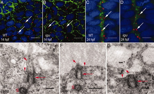
Igu is required for axonemal biogenesis, but not for the docking of basal bodies to apical membranes. A: Distribution of basal bodies (arrows) in paraxial mesodermal cells of a wild-type embryo. B: Paraxial mesodermal cells of an igu mutant embryo with wild-type pattern of basal body distribution (arrows). C: Polarized orientation of basal bodies (arrows) toward the apical membranes of cells abutting the midline of the neural tube of a wild-type embryo. D: Apical polarization of basal bodies (arrows) in the neural tube is unaffected in an igu mutant embryo. E: A representative TEM section through the neural tube of a wild-type embryo showing the basal body (long arrow) and ciliary axoneme (short arrow). The apical membrane of the ciliary pit is indicated (arrowhead). The sister centriole (S) is also visible. F,G: Similar sections through the neural tube of igu mutant embryos showing basal bodies (long arrows) apparently docked to apical membranes, but a complete lack of axonemal outgrowth (short arrows). No invagination of the apical membranes (arrowheads) to form ciliary pits is visible. In F, the sister centriole (S) is visible. In A-D, basal bodies were visualized with anti-γ-tubulin antibodies (red), cell membranes with anti-βcatenin antibodies (green) and nuclei with DAPI (blue). A and B show dorsal views of the paraxial mesodermal cells; C-G represent transverse sections through the neural tube. Scale bars = 10 μm in A,B, 5 μm in C,D, 0.2 μm in E,G, 0.5 μm in F.
|
