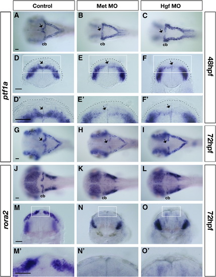
Altered expression of ventricular zone derivative markers in Met signaling morphants. Dorsal (A?C and G?L) and transverse views (D?F′ and M?O′) taken at the level of the cerebellum of control (A, D, D′, G, J, M and M′), Met morphants (B, E, E′, H, K, N and N′) and Hgf morphants (C, F, F′, I, L, O and O′) at 48 hpf (A?F′) and 72 hpf (G?O′) showing expression of ventricular zone (VZ) progenitor marker ptf1a (A?I), and of Purkinje cell marker rora2 (J?O′) in the cerebellum. At 48 hpf, ptf1a expression in the dorsal midline region of the cerebellum is missing in Met morphants (arrows in B, E and E′) and in Hgf morphants (arrows in C, F and F′) compared to controls (arrows in A, D and D′). Similarly, at 72 hpf, ptf1a expression in the dorsal midline region of the cerebellum continues to be absent in Met morphants (arrow in H) and Hgf morphants (arrow in I) compared to controls (arrow in G). In addition, at 72 hpf, expression levels of ptf1a in the rest of the cerebellum is down-regulated in Met and Hgf morphants (H and I) compared to controls (G). Neuroepithelium is outlined with a broken line in (D)?(F′). The boxed region in D?F is shown at higher magnification in (D′)?(F′). At 72 hpf, the expression of Purkinje cell marker rora2 in the entire cerebellum, including the dorsal midline region of Met morphants (K, N and N′) and Hgf morphants (L, O and O′) is significantly reduced compared to the expression in the cerebellum of control embryos (J, M and M′). The boxed region in M?O is shown at higher magnification in M′?O′. morphants, MO (morpholino) injected embryos; cb, cerebellum. Scale bar, 50 μm.
|
