
Fig. 6
- ID
- ZDB-FIG-090831-32
- Publication
- Berberoglu et al., 2009 - fezf2 expression delineates cells with proliferative potential and expressing markers of neural stem cells in the adult zebrafish brain
- Other Figures
- All Figure Page
- Back to All Figure Page
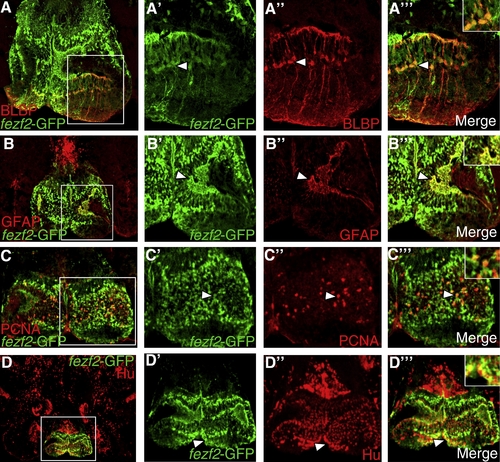
fezf2-GFP expression suggests adult neurogenesis in the caudal hypothalamus. (A) Coronal section through hypothalamus showing double-labeling of fezf2-GFP (green) and BLBP (red) (40x magnification). (A′?A″′) Closer view of the boxed region shows that some fezf2-GFP+ cells colocalize with neural stem cell marker BLBP and have radial glial morphology (arrowhead). (A″′) Inset shows colocalization in cells (depicted by arrowhead) at higher magnification. (B) Double-labeling of fezf2-GFP (green) and neural stem cell marker GFAP (red) (40x magnification). (B′?B″′) Closer view of boxed region shows colocalization in some cells (arrowhead). (B″′) Inset shows colocalization in cells at higher magnification. (C) Double-labeling of fezf2-GFP and proliferating cell marker PCNA (40x magnification). (C′?C″′) Closer view of boxed region shows colocalization in some cells (arrowhead). (C″′) Inset shows colocalization in cells at higher magnification. (D) Double-labeling of fezf2-GFP and neuronal marker Hu (20x magnification). (D′?D″′) Closer view of the boxed region shows colocalization of some fezf2-GFP+ cells with Hu (arrowhead). (D″′) Inset shows colocalization in cells at higher magnification. |
| Gene: | |
|---|---|
| Fish: | |
| Anatomical Term: | |
| Stage: | Adult |
Reprinted from Gene expression patterns : GEP, 9(6), Berberoglu, M.A., Dong, Z., Mueller, T., and Guo, S., fezf2 expression delineates cells with proliferative potential and expressing markers of neural stem cells in the adult zebrafish brain, 411-422, Copyright (2009) with permission from Elsevier. Full text @ Gene Expr. Patterns