
Fig. 5
- ID
- ZDB-FIG-090831-31
- Publication
- Berberoglu et al., 2009 - fezf2 expression delineates cells with proliferative potential and expressing markers of neural stem cells in the adult zebrafish brain
- Other Figures
- All Figure Page
- Back to All Figure Page
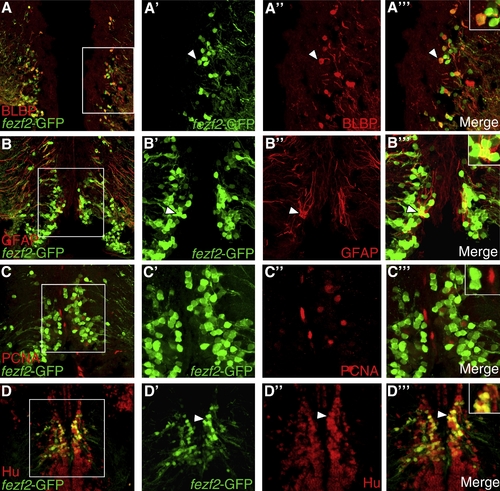
fezf2-GFP is expressed largely in postmitotic neurons of the preoptic region. (A) Coronal section through preoptic region showing double-labeling of fezf2-GFP (green) and BLBP (red) (40x magnification). (A′?A″′) Closer view of the boxed region shows that some fezf2-GFP+ cells colocalize with neural stem cell/astrocytic marker BLBP (arrowhead). (A″′) Inset shows colocalization in a single cell (depicted by arrowhead) at higher magnification. (B) Double-labeling of fezf2-GFP (green) and neural stem cell/astrocytic marker GFAP (red) (40x magnification). (B′?B″′) Closer view of boxed region shows colocalization in some cells (arrowhead). (B″′) Inset shows colocalization in some cells at higher magnification. (C) Double-labeling of fezf2-GFP and proliferating cell marker PCNA (40x magnification). (C′?C″′) Closer view of the boxed region shows that fezf2-GFP+ cells do not co-localize with PCNA. (C″′) Inset shows lack of colocalization at higher magnification. (D) Double-labeling of fezf2-GFP and neuronal marker Hu (40x magnification). (D′?D″′) Closer view of boxed region shows a number of fezf2-GFP+ cells that colocalize with Hu (arrowhead). (D″′) Inset shows colocalization in cells at higher magnification. |
| Gene: | |
|---|---|
| Fish: | |
| Anatomical Term: | |
| Stage: | Adult |
Reprinted from Gene expression patterns : GEP, 9(6), Berberoglu, M.A., Dong, Z., Mueller, T., and Guo, S., fezf2 expression delineates cells with proliferative potential and expressing markers of neural stem cells in the adult zebrafish brain, 411-422, Copyright (2009) with permission from Elsevier. Full text @ Gene Expr. Patterns